Roof Of Fourth Ventricle Anatomy

Pin By Sheena Hunter On College Brain Anatomy Medical Anatomy Medical Knowledge

Brain Atlas Of Human Anatomy With Mri Mri Brain Brain Structure Human Anatomy

14 3 Brainstem The Medulla Oblongata Relays Signals Between 1278x1500 Png Brain Anatomy Cranial Nerves Nervous System Anatomy

Midbrain5 Jpg 307 397 Basic Anatomy And Physiology Brain Anatomy Medical Anatomy

14 3 Brainstem The Medulla Oblongata Relays Signals Between The Rest Of The Brain And The Spinal Co Nervous System Anatomy Brain Anatomy Craniosacral Therapy

Brain And The Cranial Nerves Cranial Nerves Reticular Formation Nervous System

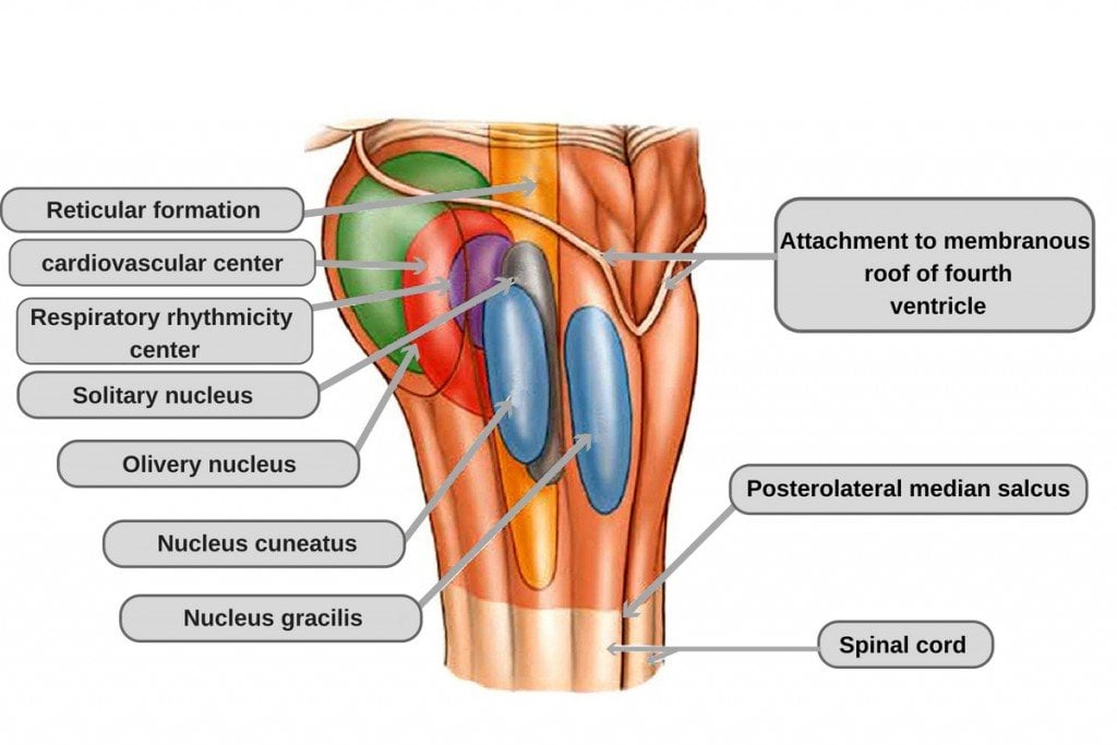
It corresponds to the ventral surface of the cerebellum.
Roof of fourth ventricle anatomy.

Mynotes4usmle Speech Language Therapy Speech And Language Speech Therapy


Cerebellar Structure Function And Vestibular Disorder Brain Anatomy Cerebellum Anatomy Human Anatomy And Physiology

Brainstem Neupsy Key In 2020 Medical School Studying Plexus Products Gross Anatomy

Cross Sectional Anatomy Of The Spinal Cord A Relationships To The Vertebra Meninges And Spinal Nerve B Ste Spinal Cord Anatomy Spinal Cord Spinal Nerve

Development Of The Spinal Cord A Early Stage Of Development B Intermediate Stage Of Development And C Late Nervous System Gross Anatomy Biology Notes

Exam 2 Chapter 14 Diagrams And Labeling Flashcards Flashcards Exam Chapter

Pin On Trauma Registrar Anatomy Pics

Protection Of The Brain Medical Massage Dura Mater Epidural Hematoma
Pin De Erendira Ruiz En Neurologia Google Imagenes Google Y Neurologia

Image Result For Facial Colliculus Facial Nerve Reticular Formation Facial

Head Ct Scan Procedure Radtechonduty Ct Scan Brain Anatomy Radiography

The Diaphragma Sellae Or Sellar Diaphragm Is The Circular Fold Of Dura Mater That Almost Completely Roofs The Fossa Hypophyseos Dura Mater Sphenoid Bone Gland

Mid Brain Structures Including Commissura Brain Structure Corpus Callosum Brain

Right Aortic Arch Double Aortic Arch And Aberrant Subclavian Artery Obgyn Key Subclavian Artery Ultrasound Ultrasound Sonography

14 3 Brainstem The Medulla Oblongata Relays Signals Between 1278x1500 Png Brain Anatomy Cranial Nerves Nervous System Anatomy

Pin By Draw It To Know It Medical On Https Drawittoknowit Com Medical School Studying Medical Knowledge Medical Terminology Study

Pin De Erendira Ruiz En Neurologia Google Imagenes Google Y Neurologia
3

Anatomy Question For Neet Pg Exam Solve This Questions For Complete Demotest Click Http Bit Ly 2hy Online Test Series Online Tests This Or That Questions

3vv Three Vessel View Diagnostic Medical Sonography Echocardiogram Medical Ultrasound

Pin On I See Sound Ob Gyn

Ependymoma Of Posterior Third Ventricle This Is A Case Of A Common Paediatric Tumour Being Found In An Uncommon Locat Radiology Radiology Imaging Mri Brain


Pin By Deb Battenfield On Radiology Notes Radiology 10 Year Old Tumor

Dandy Walker Malformation Consists Of A Group Of Anomalies Where There Is A Posterior Fossa Cyst Communicates With The Radiology Pediatric Radiology Neurology

Tencent Weibo Voice From You Echo From The World Celulas Somatico Embrion

Hypothalamic Hamartoma Radiology Reference Article Radiopaedia Org In 2020 Radiology Glial Cells Cerebral Cortex

Colloid Cyst Loc Anterior Roof Of 3rd Ventricle B W Fornices Attached To Choroid Stroma Moa Endoderm Vesigial Paraphysis Ep Cysts Brain Images Pet Ct

The Radiology Assistant Brain Tumor Systematic Approach With Images Brain Tumor Tumor Brain Images

Mri Abnormalities Following Febrile Status Epilepticus In Children The Febstat Study Http Ow Ly Dhecx Neurology Mri Status Epilepticus Medicine Book

American Journal Of Roentgenology Ajr Pagets Disease Radiography Osteoporosis

International Day Of Radiology Radiopaedia Org Neurociencia Neurologia Radiologia

3vv Three Vessel View Diagnostic Medical Sonography Echocardiogram Medical Ultrasound

Photos Of A Giant Collection Of Human Brains Human Brain Human Photographer

Color Doppler Image Choroid Plexus Papilloma Fetal Ultrasound Plexus Products

1 Chondro Osseous Junction Between The Bony Part And The Cartilaginous Part Of The Femoral Neck 2 Cartilaginous Part Of The Ultrasound Lower Limb Sonography

Oguzhan Malli Adli Kullanicinin Neurosurgery Panosundaki Pin 2020

Right Aortic Arch Double Aortic Arch And Aberrant Subclavian Artery Obgyn Key Subclavian Artery Medical Ultrasound Echocardiogram
Clear Cell Rcc Left Kidney Ctisus Com Case Study Medical Imaging Radiology

Image Result For Fetal Heart Outflow Tracts Ultrasound Obstetric Ultrasound Ultrasound Echocardiogram

Pin By N R G On Help Z Me Keep My 1st Eye Open Ascended Masters Fourth Wall Rool

Pin On Work
Https Encrypted Tbn0 Gstatic Com Images Q Tbn 3aand9gctv Bgk6epeotly Ils9h6czvsqsspbvaaly2ypwd0pnwwdcwal Usqp Cau
Source : pinterest.com
